In healthcare, as in life, no textbook lesson can provide the depth of learning that a real experience can. With today’s technology, educators can provide their students with a deeper, more realistic learning experience, especially in regards to using and studying medical imaging. Educators and students alike now have greater flexibility and a wider variety of resources available to them with the evolution of cloud-based medical imaging software. The innovative educators utilizing this technology can more easily prepare their lessons and analyze their students’ progress; not to mention, students are better equipped with the tools and experience required for their future professions.
Overview
Few people would argue that education is changing. What used to be taught from textbooks in classrooms is now being delivered through self-paced internet-based lessons. With more available resources and flexibility, online learning can be even richer in content than the same lessons delivered in a traditional classroom setting. This is true even in education around medical imaging. Whether used for advanced training for radiologists and radiology technicians or for the more general education of undergraduate biology students, medical imaging software can now deliver a uniquely detailed and accurate view into human and veterinary anatomy and physiology. This viewing experience simply isn’t possible through textbooks, basic computer-based classes, or even working on a scanning modality. As an innovative educator, a cloud-based medical imaging system can help you deliver a much deeper learning experience to your students.
Today’s educational environment requires flexibility to deliver lessons to students both within and outside the classroom asynchronously enabling self-paced learning. If you’re an educator who wants to deliver more than just two-dimensional flat images, providing access to a web-based DICOM viewer enables you to deliver a realistic experience to your students without geographic or time constraints.
Training Students
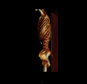
Want to show your students human or animal anatomy in three dimensions? Now you can use actual medical images displayed in a fully-functional viewer right on their la ptops. With the powerful web-based viewers available today, it’s as simple as that. Students can view color images, rotate through multiple dimensions or time, and scan through the various layers of anatomy to bring these images to life whether they’re sitting in the classroom, a clinic, or studying at home. For example, a biology professor in northern New York uses a real medical imaging system rather than a textbook to ensure his students see the same view that a doctor might when looking at human anatomy. This provides a more realistic, accurate and engaging learning experience. Not to mention, this also better prepares the students as they become more familiar with the latest medical imaging technology.
For technicians who need to hone their scanning skills, what’s better than showing them the results of their own work? With scanners ranging from radiographs to MRIs and CTs, rad techs can do their scans and then see the results of their work almost immediately on their own computer screens. A community college in Maryland uses Purview to enable students in their rad tech program to do this – immediately after doing a scan, the students can immediately view the image on their laptops and self-assess the quality of their results.
There is likely no better training for a radiologist or any other physician who will be reading studies, than the experience of viewing a medical image on an actual DICOM viewer. For these students, enabling them to view a set of patient studies so they can understand the subtle differences between diseased and normal is just what the doctor ordered.
Creating a Library of Images
For educators, it’s pretty easy to capture studies, store them and have them available for future lesson plans. Studies generated anywhere can be electronically loaded into the cloud PACS, where they can be searched and sorted as required. They can either be uploaded from any computer with a web browser interface or received electronically directly from another PACS or the modality itself.
Typically, these studies are de-identified in advance so they contain no protected health information (PHI). Often, we find that tagging the studies with DICOM tags reflecting the particular topic, modality, body part, or other identification makes presenting these to students much easier.
Developing Lesson Plans
This advanced cloud-based imaging technology also enables educators to plan lessons for their students in advance more easily. Rather than dump a term’s worth of studies on students right at the start of the semester, educators often prefer to make these studies available to them in tranches, with time-released content. This way, students are not “reading ahead” of their assignments and can focus on the current lesson topic. Lesson plans can focus on a particular modality or part of the anatomy, using corresponding medical images.
Learning Untethered
This technology also means that students are no longer restricted to the classroom in order to view their lessons. Since no specialized equipment is required, the student can use their own laptop and internet connection to view these studies in full fidelity. Classroom walls disappear and busy students can plan their work accordingly.
Students can also work through their lessons at their own pace. Those who work faster than others, will not be held back by having to wait for others to catch up. With personalized access to their online lessons, students can view images as soon as they’re ready and pace themselves through their workload.

Full Feature Viewer
With a full feature DICOM viewer at their disposal, students get the real experience of viewing and reading images; the same as a radiologist would have. The functions of the viewer are quite robust, enabling window leveling, annotations, cines, multiplanar reconstruction (MPR) and 3-D reconstructions. Just about any function the most detail-oriented radiologist would demand is available for your students. With the web-based viewer, they can learn to analyze even the subtlest conditions, annotate images with regions of interest, and export key images for additional review and scrutiny.
Add Your Guidance
Using a feature we call Study Notes, you can provide a complete set of online guidance or class notes to help direct your students’ work. Study Notes are a completely customizable document attached to the study. For example, you can pose questions to the student or give detailed direction for analyzing a specific study. With two screens, the student can view the study notes guidance on one screen and the study itself on the other. This can be the perfect way to be present without having to physically stand beside a student as they go through your lesson.
Reports from Students
If you want your students to practice giving a report of their analysis, you can have them generate their own reports in the same platform. These reports can be formatted uniquely to match your specific requirements. Once the student submits the report (either via email or in printed hard copy), you can use this to assess their capabilities and provide guidance and instruction specific to that student.
Live Assessments
Educators who wish to actively engage in the assessment of their student’s growing skills can use a collaborative function. Using this real-time collaboration, the student and the educator can both be online simultaneously, viewing the same study at the same time from different locations. This can provide the most in-depth view of the student’s navigation and analysis capabilities.
A private radiology training company in Europe uses the live collaborative assessment as the “final exam” for its radiology trainees. Near the end of their formal training, the professor joins each student online to watch and assess his or her knowledge of how to navigate, view and assess medical conditions in the provided studies.
Summary
Whether you’re training students to view human or veterinary images, using the Purview cloud-based platform to help you deliver your lessons and to assess your students can be enlightening. Lessons become more real for your students when viewing them in a medical image viewer right on their own laptops or desktop computer. Assessments can become more accurate. And your lessons can be prepared in advance, rolled out as required, and archived for future lessons. Purview enables self-paced learning and the freedom for your students to work on their lessons anywhere, anytime, on their own equipment.
About Purview
Purview enables educators around the globe to offer their students a realistic learning experience through the Purview Platform, a cloud-based medical imaging management system. By using a web-based DICOM viewer alongside a cloud PACS with controlled access, professors and students alike have great flexibility in conducting their lessons anytime, anywhere and on any device. To learn more, please visit https://www.purview.net/purview or click here to request a free, personalized demonstration!